The ASTRA toolbox: source of 5000 downloads per year, 5 patents, several industrial user licenses and a whole list of publications, citations and research partners. What began in 2008 as a small-scale postdoc experiment has grown into one of the most versatile 3D-image reconstruction platforms for X-ray tomography and other imaging techniques. Pioneers Jan Sijbers and Jan De Beenhouwer, both associated with Vision Lab (an imec research group at Antwerp University), are justifiably proud of the results already achieved and of the prospects for the future.

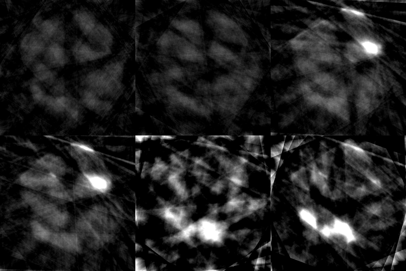
Image reconstructions of a walnut, each based on 32 scans. Left and middle via standard reconstruction algorithms. Right via neural-network algorithms.
Surfing on the success of virtual and augmented reality, 3D scans of people and objects are fairly commonplace these days. Even with just a smartphone, you go along way for certain applications. But producing a 3D scan of the inside of an object is still pretty tricky. The method most used for this is still X-rays, also known as CT scans or computer tomography. A variety of sectors already use it: inspecting luggage at the airport, quality control for products such as diamonds, fruit, flower bulbs and seeds, checking complex components in the manufacturing industry and so on. Scanning technology can offer added value in virtually every sector where visual quality inspections are used. But effectively applying this type of inspection method is not always straightforward – and that’s where the ASTRA toolbox shows its strength.
Challenges in image reconstruction
A CT scanner – the big, donut-shaped machine you have to lie in at the hospital – is probably the best-known application of computer tomography. With a CT scanner, a source of X-rays and a detector located opposite it circle round your body every few seconds to take images from various positions. Because some materials and tissues absorb the radiation more than others, this gives you contrast and hence information about the inside of your body. In the medical world, smart software then translates these two-dimensional pictures into a 3D image so that a diagnosis can be made. The technology for this standardized method in a relatively controlled environment has been operating for years.

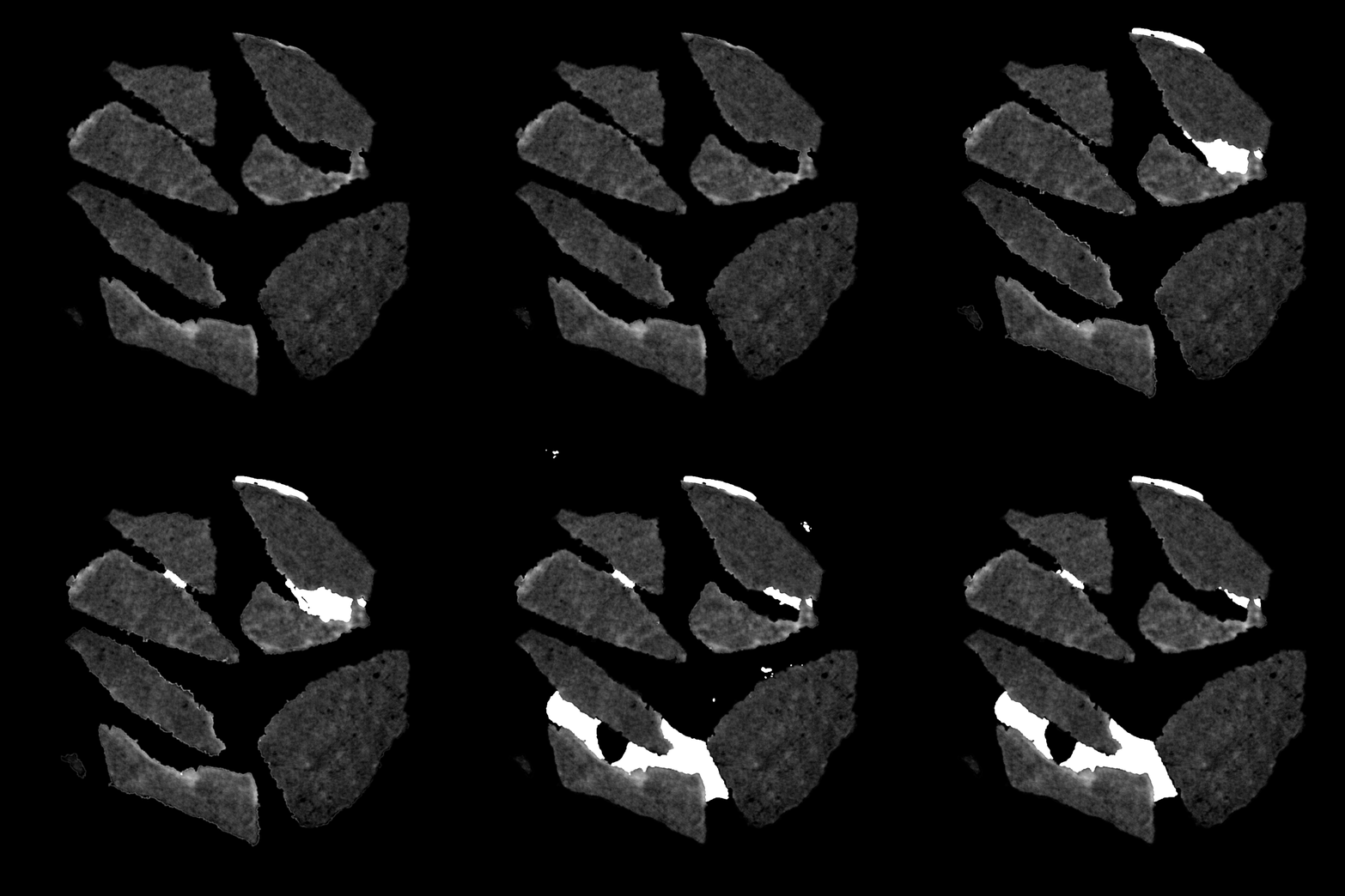
Image reconstructions of a plastic product part, showing its internal cracks. Top images reconstructed via standard algorithms need 1500 scans. Bottom images made by neural-network algorithms only need 50 scans.
But what happens if the circumstances are not ideal? For example if the scanner is unable to rotate fully round the object? Or if it takes a track other than a true circle? Sometimes certain materials can cause interference. If the object moves, there can be random noise or unclear images. In some cases you may want to limit the number of two-dimensional images to a minimum for health reasons (X-rays are harmful for humans) or to save time and money. Ten years ago, there was no software at all to perform image reconstruction in this context. Which is why, in 2008, Vision Lab began developing the ASTRA toolbox. In the early years, Vision Lab received support from the Flemish Innovation and Enterprise Agency (VLAIO) and since 2014, development has been in conjunction with CWI, the Centre for Mathematics and Computing in Amsterdam.
 |  |
4D image reconstruction of a liquid flowing through rocks. Left: generic algorithms show high noise levels and interference from the moving liquid. Right: smart algorithms deliver a high-quality result based on the same number of scans.
Future-proof
The greatest strength of the ASTRA toolbox is that its development always stays abreast of important trends and challenges. One of these is 4D imaging, in which objects change over a specific period of time. This is one of the major focal points in current academic research. For example, how do you produce an image of a continuous flow of liquid, such as the contrast agent used in a medical scan? Or the compression of foam? And what do you do with objects passing along a conveyor belt and you don’t know what position to ‘photograph’ them in? Vision Lab has developed prototype algorithms for all of these scenarios – some based on neural networks and self-learning systems – which can be programmed as an application layer on top of the generic source code of the toolbox.


4D image reconstruction of a foam being compressed. Both images are based on the same number of scans. Top-image reconstruction algorithms treat each scan individually. Bottom image shows higher resolution and less noise because the reconstruction algorithm correlates each scan with the information of the previous ones to extract additional information on the foam's behavior.
One of Vision Lab’s other areas of development is imaging based on phase shift instead of attenuation. In certain cases, this technology delivers greater contrast and hence more details. Vision Lab is also doing pioneering work here on compatibility with flexible and non-standard setups.
In addition, the toolbox can be used for image reconstruction via other electromagnetic sources. This might be electron microscopy or multispectral imaging. Vision Lab is also working with imec Florida on Terahertz Imaging, a technology that uses non-harmful radiation and which could well bring about a revolution in (medical) imaging.
Finally, Vision Lab is working on hardware solutions aiming to achieve the same level of flexibility they have reached in the ASTRA toolbox software. For example, by allowing three degrees of freedom in the configuration of source, sample and detector and by establishing a smart link with the software, it is possible to obtain an excellent result in virtually every scenario with a minimum number of images.
Versatile and flexible
In the meantime, the most versatile software package has already been online for over six years. For the experts among us: the current version supports supports 2D parallel and fan beam geometries, and 3D parallel and cone beam. All of them have highly flexible source/detector positioning. A large number of 2D and 3D algorithms are available, including FBP, SIRT, SART, CGLS. Selecting the right one guarantees optimum results. All basic forward and backward projection operations are GPU-accelerated and can be retrieved directly and adjusted using the Python or Matlab programs. For those who find this all sounds a bit like gibberish, Vision Lab runs training courses and offers technical support to research partners and paying customers.
The Vision Lab software package is downloaded about 5000 times each year. Users tend to come from two major segments: university research groups worldwide, who use the package for applications in medicine and physics, as well as for imaging techniques. For example, the software runs in a number of particle accelerators, such as the ESRF in Grenoble, where materials are being researched using the most intense X-ray detector in the world. The second target group is companies: for example operating in the energy, food and automobile industry or for inspecting diamonds, 3D prints and complex quality products. Thanks to the speed and versatility of the software, the ASTRA toolbox has become a must-have attribute in many industries and sectors.

Left side of the arrow: image reconstruction of a gold nanoparticle. Only greyscale information can be extracted. Right side of the arrow: thanks to smart algorithms, renderings can be made of individual atoms allowing to analyze the crystal structure.
Open source and valorization
Anyone with sufficient expertise can download the source code for the ASTRA toolbox via GitHub. Vision Lab made a conscious choice from the start to share the generic code as open source. This decision has stimulated its research significantly, including a sharp rise in the number of users. It also helped the research group itself to quickly gain recognition and relevance in its sector. When it becomes clear that you have nothing to hide, other organizations are usually willing to open their doors more quickly to you. And the developers at Vision Lab have received many credits (publications, citations…) in return for what they have made available. Finally, it is a sign of quality when many users are able to download and implement your software without any problems.
All this now opens the way to further valorization. The generic source code is fully online, but imec Vision Lab has also developed a number of specific industrial applications. Imec Vision Lab is reaching out to companies that want to use their technology to increase the quality and efficiency of their production process. Companies are not always aware of the opportunities provided by the technology and often come to realize that its potential is far greater than they had first thought.
Want to know more?
- The ASTRA toolbox download and documentation is available free of charge online.

Jan Sijbers graduated in Physics in 1993. In 1998, he received a PhD in Physics from the University of Antwerp, entitled Signal and Noise Estimation from Magnetic Resonance Images, for which he received the Scientific Award BARCO NV in 1999. He was an FWO Postdoc at the University of Antwerp and the Delft University of Technology from 2002-2008. In 2010, he was appointed as a senior lecturer at the University of Antwerp. In 2014, he became a full professor. Jan Sijbers has published 250+ journal papers and is Associate Editor of IEEE Transactions on Image Processing as well as of IEEE Transactions on Medical Imaging. He is the head of imec-Vision Lab and co-founder of IcoMetrix.

Jan De Beenhouwer received an MSc in Computer Science Engineering in 2003 from KU Leuven, Belgium and a PhD in Biomedical Engineering from the University of Ghent, Belgium in 2008. He was a postdoctoral fellow for 2 years at the same institution prior to joining the Vision Lab at the University of Antwerp, Belgium, where he is now active as a research leader of the ASTRA tomography group. His main interest is in image reconstruction focusing on X-ray computed tomography and electron tomography.
More about these topics:
Published on:
2 September 2018


